We modern homemakers do not have the Exact Same sixth sense with cleaning Solutions our grandparents formerly cultivated. While they can get rid of any stain or dissipate any odor equipped with only a toothbrush and a few tricky soap, then we have a tendency to favor the trusty brand name cleansers that do just what they say. After all, what is the purpose of pouring baking soda to your fatty oven only to wind up with a white, powdery residue you are going to need to wash up for months? While our reluctance is clear, it turns out that not every wacky suggestion you discover on the world wide web is a flop -- even a few weirdly magic cleaning solutions from cleaning services Ashburn, VA really get the work done better and quicker than their chemical choices.
Applying ingredients you have already got on your pantry, handle those tough jumble Areas in your house that are not getting their cleaning needs fulfilled by sprays and exfoliates.
If your linens or towels are starting to Develop a mould stench, provide them a refreshing scrub in a vinegar bath. Your weary textiles will appear entirely fresh-scented and re-fluffed, without a hint of the dank odor. Save laundry detergent, and delight in this age-old solution.
Eliminate rings on timber with olive oil and salt
Though your coffee table is Beginning to show the usage of coaster-less cups and cups, do not begin searching for a replacement just yet! This kitchen mix is just the thing to facilitate those stains from your grain and also receive your wooden surfaces back to their own glistening origins. Do not get frustrated if the rings are still there once you wash off your mix -- only check back afterwards and revel in the vanishing act.
You understand that some ovens are Self-cleaning, but what is this about microwaves? It is true -- you can allow your microwave do the task for you personally by heating some water and lemon juice or vinegar at an microwave-safe bowl for 5 minutes, allowing the mixture sit cool, then wiping the sides down. Any built-up dirt and dirt will probably likely be long gone, with no you out yourself.
Rub silver away stinks with toothpaste
Rather than the standard smelly silver Cleansers, try out this minty-fresh alternate. Simply apply toothpaste for your Bits and rub it with a rag. to get Into any cracks or elaborate designs.
When a home is available, the Objective is to make it as appealing as possible To prospective buyers. The very cost-efficient method to liven up the house is to wash it completely.
Eliminating Clutter
Many prospective buyers might not see beyond clutter and dirt. If the home is Packed with the proprietor's"stuff," they'll assume that the home is too little for their own possessions. Too much mess also presents a security - and - purchasing - hazard! Someone who trips items left on the staircase will probably have a fantastic feeling about your home. Moreover, a sterile, clutter-free surroundings is much more appealing than a filthy, one.
Items which are no longer desired. Matters which are in good shape can be donated or sold to some charity. Rooms will look more spacious, cabinets will appear larger and shelves will appear roomier to prospective buyers.
Time to Wash
Professionals from maid services montgomery advice to concentrate on the rooms which prospective buyers are interested in - dwelling Room, dining area, kitchen and bath. Look at each area as a possible buyer may and see what catches the attention. Ensure the starting point for your cleanup procedure.
Give significant focus on the kitchen and baths. Cleaning products to be certain they're acceptable for the surfaces to be washed. Abrasive cleansers deliver more cleaning power for hard-to-remove lands such as food particles and dirt residue in bathrooms. But they could be overly unpleasant for surfaces which may be scratched, such as laminate or solid surface counter tops. Generally, gel and liquid cleansers tend to be somewhat less abrasive than powders.
Spray cleaners are Simple to use for small regions, like countertops, while Powders or fluids mixed in a pail of water tend to be somewhat more efficient for larger regions, like walls and flooring. Flooring can get cloudy from cleaning solution residue, which makes them seem dirty when they are actually clean. To avoid this, use a no-rinse merchandise or wash the ground well after every cleaning.
Mold and mildew are particularly problematic in toilets. With all the Publicity regarding the disorders that mould can cause, potential property buyers are more sensitive than ever about its existence.
Utilize a non-streaking cleaning merchandise, like a glass or glass and A normal routine, such as draining the bathtub after washing, with a little squeegee on shower walls after every usage, and drying taps and manages to reduce water stains, will keep things tidy. Use a dusting product so the dust will be trapped and eliminated instead of dispersed in the atmosphere. Vacuum frequently, with long straight strokes.
If repainting isn't from the budget, carefully wash surfaces to remove dust, fingerprints and dirt. Painted surfaces are often washable, but try the cleaning solution in an inconspicuous place first. Utilize a non-abrasive, cleaner. To prevent chain marks when cleaning big vertical places, begin at the bottom and work upward, overlapping regions and having a circular movement.
Keeping along with these cleaning chores will probably make your house look its very best. And, once a purchaser is found, it is going to be simple to get the house"broom ready" for this last walk-through before closure day.
Away With Clutter! This way, in case an unexpected buyer falls in, the home will be prepared to show.
Clear off the kitchen and bath counters, sorting and saving Wipe surfaces clean. Whatever will return on the countertop ought to be wiped clean, also. From the kitchen, keep outside just those appliances which are used every couple of days.
Do not let papers and email pile up. Sort Daily and discard what is not needed. Put a couple baskets in strategic places around the home. When the realtor requires, the clutter that's an inevitable part of everyday life could be immediately scooped up in the baskets and hidden off.
Selling Your House?
Here are some daily, Fast cleaning hints for when prospective buyers make spur Of the minute visits:
Wipe Away. Use disposable wipes to quick-clean bathroom bowls and regions from the kitchen.
Scented Toilet. Toilet bowls every evening before bed. At the morning, each toilet will smell great!
Switch It On. Shower, therefore walls remain bright and clean without additional cleaning.
Dish DOs. Water and simmer until you prepare a meal. Insert Huge pots And pans that will not fit in the dishwasher since you use them. From the time
Require Two. If you reside in a two-story home, do not eliminate your old vacuumcleaner. Maintain it upstairs so that you do not need to drag it up and down stairs. It will Save you energy and time. Floor of your property.
Sterile As You Move! This will make demanding jobs a lot Simpler.
Carpets to the Rescue! Use mats or rugs at all entrances to capture grit and dirt that Can develop on flooring and carpets.
You many not know it, but your home is hazardous to your well being. Insect Droppings, dust mites, bacteria-laden sponges, spoiled food - all lead to a myriad of health problems which range from asthma and allergies to gastrointestinal upsets. Additionally, many cleaning products include volatile organic chemicals (VOLs), which pose several health risks. There's a solution. Here is what cleaning services in Springboro suggest.
Sterile in an Organized Manner
There is no point in cleansing the floor simply to dust the ceiling fan and Deposit a gray movie over everything again. To wash nicely -- which means to wash healthily -- you want to wash efficiently, preventing going back and forth about a space. Rather, work employing a systematic strategy. Think in relation to left to right, top to bottom. Begin with walls and ceilings, and work your way down to furniture and windows, finishing with all the flooring.
Sterile Things You'd Never Think to sterile Lay the mattress cover in very hot water (60 degrees C or longer) monthly, and wiping the cover of the mattress with warm water, can go a long way towards reducing morning stiffness.
Infection With Old Wool Clothing
Wool creates static when rubbed onto a surface. Furniture dust-free with no spray or gloss. hazardous ingredients. Rather, dab toothpaste with your palms or rub it on with a cloth. For bigger bowls and trays, use a glue made from bicarbonate soda mixed with water onto a moist sponge.
Clean Your Drains that the Non-Toxic Method
Chemical drain cleaners are extremely corrosive and harmful, including Such noxious ingredients like lye or malic acid. The vapours are detrimental. Rather pour a kettle of boiling water or throw a few of bicarbonate followed Also effective, especially in Preventing blockages, are lots of brands of enzymatic cleaners located in pet stores.
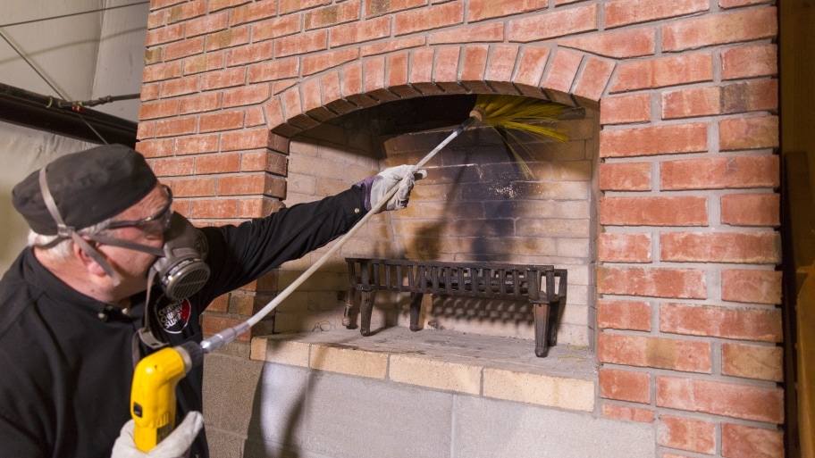
It takes only 15 minutes for gas fireplaces columbus, oh cleaning. Here is what you will want: a kneeling pad, a scoop and brush set, a wash brush, glass cleaner, an apron, a huge plastic sheeting, rubber gloves, and a lidded garbage can, used coffee grounds, a spray bottle full of water, dishwashing liquid, and a soft fabric, and paper towels.
Wait at least 24 hours once you have burnt your final fire so that the ashes cool completely.
Wear an apron and put a plastic sheeting in the front of the fireplace.
Maintain burnable logs; throw badly charred logs to the garbage.
Sprinkle a few of used coffee grounds to the ash to minimize flyaways. Sweep each inside wall top to bottom using all the fireplace brush. Shovel the ashes to a pan, then ditch the debris to the garbage.
Sweep each display top to bottom using all the fireplace brush.
Tackle the outside. For brick: Spray water on sooty regions followed by hearth cleaner. (For bricks over 50 years old, use just waterwithout a hearth cleaner.) For stone and marble: Spray residue with warm water, wash with dishwashing liquid and a fabric, rinse, and dry.
If the fireplace has glass doors, then employ hearth cleaner into the rear and front of every doorway, then wipe down a single panel at a time, eliminating the cleaner until it dries.
Alter the facsimile and grate. Carefully match and throw the tarp. Pick out the garbage can out. Now draw the displays, shut the doors, and reunite. But do not create a flame just yet. You should like that sparkling-new fireplace for at least a couple of days, right?
Professional secrets from Office cleaning Durham NC that may make your home sparkle: Remove Bathing room Soap Scum Soap includes a nasty method of forming a hard-to-get rid of film on tile inside showers and tubs. You won't eliminate it by rubbing. Rather, wait for the top to dry, scrape off the scum with a 4-in then. plastic material putty knife. For grout ranges and textured surfaces, work with a Mr. Clean Miracle Eraser. To avoid soap scum buildup, cease using actual soap and start using a man made. Chemically talking, any soap in a gel or liquid form, plus some bar soaps (Zest and Ivory), are in fact synthetic soaps and far less inclined to leave a hardcore film in your sink, tub or shower. Scum-Proof Your Shower Doors Keeping shower doors clear and streak free is really a challenge-unless you understand the pros' secrets. Begin by cleansing any mold, streaks or mildew off the cup with a cup cleaner. Work with a Mr. Clean Miracle Eraser to find yourself in the cracks in textured cup. Scrape off difficult buildup with a razor blade. Dry the hinged doorways with a cloth. Treat the doorways with something like Rain-X. These glass treatments type a low profile film on the cup to improve water repellency, causing soap and drinking water to bead up and elope the glass. (Squeegee off the drinking water after bathing to help keep soap scum from accumulating again.) Spray or clean on the glass therapy, wipe it off with a new microfiber cloth then. Overspray won't damage surrounding surfaces. The merchandise repel water for half a year. Trash Can Cleanup Use a liquid toilet pan cleaner to clean the inside of a new dirty trash can. It'll cling to the relative sides for better cleaning. A toilet brush will inside help you reach down. Rinse nicely for a clear can. Duster for the Vertically Challenged Unless you perform in the NBA, dusting fans along with other high, out-of-reach objects is a true chore. Wrap a dryer sheet around a clear painting roller and safe the ends with elastic bands. Attach an extension handle to the dust and roller away. Shop Vacuum cleaner Mop Bucket The dust collection portion of your shop vacuum can make a slick-rolling bucket for mop water. It is possible to load it with sudsy drinking water and work quick and effectively-no lugging much bucket around again. And when you're carried out mopping, simply roll it to the sink or ground drain to empty.
After fire damage it truly is natural to need to jump best in and clean the contents and creating. Action is actually a great assist timely, but incorrect activities can jeopardize or impede satisfactory restoration.
Crystal clear and protect chrome trim about faucets and also other bright function by cleansing with detergent and utilizing a covering of Vaseline or gas.
Blow off or even brush-vacuum loose smoke cigarettes contaminants from upholstery, draperies and carpeting.
Open glass windows for ventilation if weather premits.
Empty freezers and refrigerators if energy is off, and prop doors start with a rolled towel or newspaper allowing air to circulate.
Pour antifreeze inside of toilet bowls, tanks, sink and tub drains in order to avoid freeze harm if warmth is off inside the wintertime.
Call a fresh plumber in order to drain and blow out there all normal water lines in case warmth is off in the winter.
Remove pets to clean environment if weighty 1st residues are available completely.
Send an example band of garments for cleaning and deodorization in order to observe the total results.
Retain a fresh contractor to desk up open glass windows, roofs, or various other penetrations in order to prevent additional damage.
Do Not:
Wipe or make an effort to clear fire residues from surfaces, ceilings, or various other absorbent surfaces.
Take advantage of carpeting or even upholstered furnishings influenced by weighty smoke residues or even particles.
Use food items or canned goods put through heat.
Turn on personal computers, televisions, stereos or even electrical kitchen appliances until they're checked and cleaned.
Clear and gently sanitize your wood chopping blocks and trimming boards by rubbing with a lemon or spritzing them with homemade bleach. If residue continues to be from the lemon fruit juice, wash it off following the wood is dry completely.
To clean the within of grimy bottles and glassware which are difficult to completely clean (say, a bottle of wine that’s been used mainly because a flower vase or perhaps a narrow-mouthed mason jar following a long fermentation) team of cleaning professionals from themaidsofcincy.com/ recommend to utilize uncooked rice. Sprinkle 1/4 mug of uncooked rice along with a several tablespoons of drinking water and a little squirt of dish soap in to the bottle, cap and swish before bottle is clean after that.
To polish stainless silverware and get gone the tarnished appear that builds-up as time passes (especially if you utilize a dishwasher instead of hand-wash), rub each utensil with a solid baking soda paste, then wash and dry well.
And talking of stainless, rub simple baking soda or our nontoxic kitchen cleanser all around the inside of your stainless sinks at least one time a week to help keep them spot-free of charge and gleaming.
Descale kettles by filling them with equivalent parts whitened vinegar and water (make use of distilled water in the event that you live in a location with hard plain tap water), bring to a new boil, then permit sit for a number of hours or overnight. Rinse well, after that boil once with simple water to eliminate any lingering vinegar residue.
To eliminate the stubborn gunk about non-porous surfaces (like the stove top), create a paste away of baking soda and hydrogen peroxide to scrub those stubborn splatters away. Let it sit for some moments before scrubbing, if required.
Work with a toothbrush or utilized dental care Sulcabrush to scrub walls splash tiles, grout, caulking, any crannies within your fridge, and baseboards, especially near any tables or areas where food is served or consumed often.
Dust allergies ensure it is difficult to breathe and could trigger asthma symptoms also, such as for example wheezing, coughing, tightness in the shortness and upper body of breath.
Dust also just itchy makes some people.
People with dust allergy symptoms often suffer probably the most of their own homes or even in other people’s houses. Oddly enough, their signs and symptoms worsen during or soon after vacuuming frequently, sweeping and dusting. The procedure of cleaning can mix up dirt particles, making them better to inhale.
Dust Allergy Symptoms
Sneezing
Stuffy or runny nose
Crimson, itchy or teary eyes
Wheezing, coughing, tightness in the upper body and shortness of breath
Itching
Dust Allergy Triggers
Dust mites
Cockroaches
Mold
Pollen
Pet hair, feathers or fur
Dust Allergy Triggers
Dust mites. Dirt mites-sometimes called mattress mites-are the most typical reason behind allergy from house dirt. Dust mites live and multiply very easily in warm, humid locations. They prefer temps at or above 70 degrees Fahrenheit with humidity of 75 to 80 percent. They die once the humidity drops below 50 percent. They're not within dry climates usually.
Dust mite particles are found in pillows, mattresses, carpeting and upholstered furnishings. They float in to the air flow when anyone vacuums, walks on a disturbs or carpet bedding plus they settle after the disturbance is over.
Dust mites certainly are a common reason behind asthma in children.
A house doesn't need to be dirty to trigger a dirt mite allergy reaction visibly. The particles are too tiny to be observed and can't be removed using normal cleaning procedures often. Actually, a vigorous cleaning could make an allergic person’s signs and symptoms worse.
Cockroaches. Cockroaches reside in all types of neighborhoods and structures. Some social people develop allergic reactions when they remain cockroaches. Tiny contaminants from the cockroach certainly are a common element of household dust and could be the true reason behind a dust allergy.
Mold. Mold is really a fungus which makes spores that float in the air. When people who have a mold allergy inhale the spores, they obtain allergy symptoms. There are various kinds of mold-some types you can observe, others you can’t.
Molds live everywhere-on logs and on fallen results in, and in moist locations like bathrooms and kitchens. Tiny mold contaminants and spores certainly are a common element of household dust and could be the true reason behind a dust allergy.
Pollen. Pollen originates from trees, grasses, weeds and flowers. People could be allergic to various kinds of pollen. For example, some social people are allergic to pollen from just beech trees; others are usually allergic to pollen from just certain forms of grasses. Pollen is really a common element of household dust and could be the true reason behind a dust allergy.
Animal hair, feathers and fur. Pets can cause issues for allergic individuals in a number of ways. Their dander (pores and skin flakes), urine and saliva could cause an allergic reaction, when coupled with household dust especially. In households with birds, feathers and bird droppings may also turn out to be embedded in home dust and cause issues for those who are allergic to them
From clearing up broken cup to deterring pests or tackling stains on furniture and clothing, top cleaning company in central maryland suggested simple, organic solutions that work.
Eucalyptus oil gets rid of the gummy residue still left by shop stickers.
Buffing the marble tabletop with vehicle polish leaves a slim, invisible film that assists decrease the risk of stains.
To eliminate furniture indentations from pure wool carpeting location a tea towel on the area and press with a warm iron. Heat shall lift the fibres. Do not try this with artificial or perhaps a wool/synthetic mix carpet.
Light a match up and allow it burn a couple of seconds to eliminate toilet smells.
To avoid bathroom mirrors steaming upward, regularly rub a dried out bar of soap on the surface area and rub in with a clean cloth.
Stop clothing with thin straps dropping off hangers simply by sticking small felt furnishings pads onto the hanger only beyond where in fact the straps sit.
To eliminate oil from silk clothing, rub cornflour in to the region and lightly brush off gently. Cover the oil tag completely with an increase of cornflour and keep to sit for some hours. Shake clothing free from flour and hand wash, or work with a gentle machine period, using soap ideal for delicates.
To help keep spiders or any nasty surprises away of shoes you retain outside, (such as for example your gardening shoes or function boots), location old stockings outrageous of them. Make certain the stockings have holes inside them don’t, and when they don’t match snugly outrageous, use an rubber band to secure them.
To make candles go longer, cover with a plastic place and handbag in the freezer every day and night before lighting.
To keep your vehicle windows frost and ice totally free when left outdoors overnight within the wintertime, mix three parts vinegar to 1 part drinking water, put it within a spray bottle and spray about the windows mainly because needed.
To prevent control keys from becoming undone or reduce, dab a little very clear nail varnish at the top thread or onto the stem of the thread and keep to dry.
To eliminate pollen from the stamen of plants, take a little bit of sticky tape about five centimetres very long, gently push the sticky part to the pollen tag and lift off. Do it again with clear sticky tape as needed. Do not make an effort to brush it off.
To reuse the items of soap that are left over always, mix them with glycerine plus some warm drinking water. Pour right into a bottle for a handmade liquid soap.
To pick up little fragments of broken cup, press pieces of breads onto the affected area.
If an aquarium is had by you, save the water each right time you change it out and water your home plants with it. It’s filled with nutrients and makes an excellent fertiliser.
To avoid ants entering your home, draw a chalk collection on the floor where you need them to stop. If you reside in a rainy region where ants certainly are a problem, you need to re-draw the chalk ranges each right time it rains.
To deter silverfish, location entire cloves in wardrobes and drawers.
To obtain blood out of materials, use hydrogen peroxide. Apply it right to the stain and launder in the washer.
To remove body essential oil stains from cuffs and collars of coloured t-shirts and blouses, rub hair shampoo on the stains. Rinse out the hair shampoo, then wash the clothing as usual.
To regenerate a vase of wilted plants, put in a teaspoon of mild detergent.
Make use of leftover styrofoam peanuts while great drainage in underneath of a pot.
To avoid drawers from sticking, rub a bar of soap over the runners to create them glide smoothly.
To avoid ash from flying when cleaning up a fireplace everywhere, work with a spray bottle filled up with water to cover up the ashes with a lighting mist.
To clean underneath of the iron, sprinkle salt about the ironing table and iron backwards and forwards.
To find light switches at night, put a dot of luminous color on tape and adhere to the switches.
To help keep pinking shears or scissors sharp, cut by way of a sheet of folded aluminium foil or coarse sandpaper.
To leave an available room smelling fresh once you have vacuumed, place several drops of one's favourite gas (such as for example lavender or peppermint) close to the vent where the heat is released. The air warms the essential oil and blows it in to the room.
To mask unpleasant odors, put some coffees in a saucepan and burn off them. The odor of espresso shall overpower another nasty odors.
To completely clean a microwave oven, put four tablespoons of lemon juice to 1 cup of water within a microwave-safe and sound bowl. Boil for 5 minutes in the microwave, permitting the steam to condense inside wall space of the oven. After that clean them with a smooth cloth.
To completely clean a stainless-metal sink, put the stopper within the sink with two denture-cleaning pills and half fill with drinking water; leave for a number of hours or over night and the very next day it must be sparkling. Use the water to completely clean the draining board then, too.
To eliminate fingerprints from stainless-steel home appliances, place handful of baby oil about a napkin and wipe the impacted areas. The fingerprints will wipe away just.
To eliminate marker pen off really hard surfaces, spray on tresses spray and wipe it off.
To eliminate cat and dog tresses from clothes and furniture, rub them with damp rubber gloves.
With regards to bathroom maintenance, it isn’t the most attractive facet of apartment living, could it be? Actually, it can sometimes be quite disgusting, depending on who you're living with aswell. You can encounter overflowing toilets, clogged drains, unclean bath mats and sluggish plumbing - which can easily create a large amount of mess and problems for apartment tenant.
Although your landlord is in charge of major repairs, maintaining your bathroom maintained and clean can go quite a distance to avoid annoying bathroom problems to cope with.
So check out quite a few helpful practical bathroom maintenance and cleaning suggestions from the largest selection of housekeeping services we've listed below so that you can like a clean, worry totally free bathroom. What you should do is:
· Make sure to completely clean the toilet regularly
Obviously we find out that may not be the almost all pleasant of household chores, nevertheless your toilet sees a complete lot of action every day, therefore cleaning it is a must properly. So, though disinfecting the bathroom . bowl ought to be a no-brainer even, cleaning the base of one's toilet is as important because it may also collect germs and germs just.
· Antimicrobial bathroom mat
You’ll be surprised to start to see the huge distinction this type of mat does within your bathrooms. If you are fighting bad bathroom odor, pee stains on to the floor, germs spreading, this antimicrobial mat is crucial for the bathroom floor then. It’ll keep your bathrooms fresh and clear and germ free.
· Mirrors and cabinets want cleaning, too
Toothpaste splatter, hair items, and daily starting and closing may foster lots of germs and bacteria on your own bathroom cupboards and mirrors. So, be sure to maintain an all-objective cleaner beneath the sink for the casual wipe-down.
· Invest inside antibacterial wipes for the sink
Makeup, dirty hands, tresses and a number of clog-exacerbating items help to make their way in to the sink every full day. Not merely do antibacterial wipes maintain these items from your drains-they are wonderful to possess in a pinch when unpredicted guests show up. It takes merely in regards to a minute to clear your sink with one of these handy wipes.
· Clean the toilet floor
Just think about any of it: your dried out feet and shoes drag within dirt within from the fantastic outdoors, as well as your wet feet track moisture onto the ground after a shower, therefore it is practical that lots of bathroom floors could be filthy surprisingly. Being a high-traffic region, it’s far better sweep and mop your bathrooms floor at least one time a week, and even more when you have children or pets maybe.
· Handling Rust
Probably the most irritating areas of bathroom maintenance may be the collection of corrosion that inevitably happens. Therefore numerous toiletries leave an urgent rust ring; but an instant spritz the correct cleaning products will need the rust quickly. If you are concerned about harmful chemicals, then simply pour salt on half a rub and lemon it on the stain for green cleaning.
· Clogs - a standard problem
Most people who've lived within an apartment have handled clogs at least one time. Rather than running to the shop to obtain a pricey item to unclog your drain, open your kitchen area cabinet and make your personal product just. Mix 1 mug of baking soda with 1 mug of salt and ¼ mug of lotion of tartar. Maintain this mixture within an airtight container and pour about ½ cup of the substance straight down the drain every couple of weeks, accompanied by a quart of boiling drinking water. You’ll see that is an easy, natural way to maintain your drains clog-free.
While you’re preparing your closet and home for warmer weather, be sure to examine these springtime cleaning tips distributed by regular house cleaning services:
Clean Based on YOUR TIME AND EFFORT Constraints
In the event that you don’t have a complete lot of time, it’s far better complete one job at the same time rather than attempting to work on a number of things at once.
Handle smaller jobs like dusting when you're able to, and tackle your closet if you have a small more time and energy to do laundry, finish off your winter clothing, and grab your shorts.
Start with De-Cluttering
Our homes, our bathrooms particularly, are filled with items we don’t make use of. Go through your discard and bathroom expired cosmetics and cosmetics. It’s also smart to replace batteries and up-date your medical kits. In case you have children, it’s likely that you’ll be making use of first aids kits much more in the coming weeks.
You should check out items around your complete home also. If something is not any functional longer, re-purpose it or take it off. Utilize this as a right time for thrift shop donations or hand-me-downs.
Utilize the Right Tools
You want your home to look as if you spent hours scrubbing it, nevertheless, you can clean faster than you imagine. Several products offer dirt- and grime-attracting disposable microfiber cloths to create your life easier. They’re inexpensive and an easy task to discard when you’re done with them generally.
A good vacuum, vacuum pressure with HEPA filtration especially, will ensure all of the dust and undesirable particles are taken off your floors. These full days vacuums include various helpful accessories just forget about stairs, canine beds, ceilings, fans, and much more. Those small tools can make a difference really. Cordless vacuums are becoming popular since they’re lightweight also, need an outlet don’t, and can reach about anywhere just.
You may also desire to contemplate using a fan once you don’t need AC or even heat. It shall help keep your air flow from getting stagnant.
Work throughout always
This can force dirt downward and keep you from needing to re-dust or re-clean your space. In case you have a vacuum with an extended extension hose, use it to obtain dust and cobwebs from your own ceilings and fans. Then dust your furnishings and other products before vacuuming all of the dust from your own floors.
Let Spring Cleaning Collection a New Tone
If your space feels heavy and dark, do things that help to make it airy and light. If you’re sick and tired of the bland uniform wall space, add new vibrant art or pillows to improve the space. Replacing stuff like bedding, towels, desk linens, and window treatments can be inexpensive methods to transform your space even.
REQUIRE Help When Needed
Along the way of cleaning, you might discover bigger or tougher jobs than you're expecting. Should this happen, don’t be scared to hire an inexpensive handyman or ask an experienced friend for assistance.
No matter the method that you intend to tackle your springtime cleaning, remember it’s made to tidy your area and plan a fresh year. You should undertake it with some notion of the method that you want your area to check and feel as soon as you’re done.
Start with a clear dishwasher and a clear sink. This will be such a very simple, but exclusive cleaning for health suggestion. Ensure that your dishwasher as well as your sink are usually empty before you begin cooking; that way, it is possible to wash and load messy equipment and dishes immediately rather than leaving them to sit down out for a couple hours (or immediately, or days!).
Clean as you set off. Having an empty sink and dishwasher in the beginning of one's meal prep, cleaning as you go gets easier!
Do quite a few maintenance work monthly on your own knives and cookware. Have to clean burnt-on staining and remove rust places from your own stainless steel pans and pots? Or just remove staining and polish? Your knives possess gotten just a little rusty or spotty maybe, and you need to get them fresh and shiny again, or they have to be honed. Monthly it's wise to take share of the items and tackle any problems before they escape control and have a whole afternoon to handle.
Oil your trimming boards monthly. On a single note, all it requires is 5 minutes per month to keep up your wooden trimming boards. You can do this for the wooden spoons also! This saves money and time; you'll replace these things much less frequently if they're cared for.
Obtain the right cleaning equipment, and keep them near by. There's nothing even more frustrating than on the point of do a small cleaning or maintenance function and then be thwarted as you either a) you do not have the right device or, b) you need to run around your home finding it. However, you almost certainly don't need 10 various kitchen area cleaners clogging up your under-sink space. Pare right down to the essentials and store them neatly beneath the sink then, either in a pull-out drawer, some buckets and baskets, or within an over-the-cabinet door rack.
Keep your countertops clean, and clean them properly. Messy countertops - heck, countertops which have pretty things even, but too many of them just! - could make a kitchen area sense untidy and claustrophobic, whereas mostly obvious countertops are usually invigorating and inspiring. They invite one to cook also, rather than feeling like your kitchen area is unready for you personally. Try clearing a couple of things off your counter top, and you will see why!
Bid farewell to cleaners that want rubber gloves and a new gas mask, and state hello to great old-fashioned healthy household cleaning solutions that the job equally well. It is possible to thank your granny for them.
While we’ve produced some really welcome advances in the wonderful world of domestic goddessness (many thanks from underneath of my coronary heart, oh washer inventor), there are several things that in no way needed improving upon perhaps. With regards to household cleansing, eschewing harsh chemical substances and cleaning brokers and opting rather for all those good old-fashioned cleansing remedies of yesteryear will be kinder to your bodies, kinder to the surroundings and usually kinder to your bank balance too! Have a look at these 20 easy and natural answers to your cleaning problems.
Shampoo for the sofa
Whoever has kids can reap the benefits of this upholstery hair shampoo for furniture fabric. Blend 6 tablespoons of real soap flakes and 2 tablespoons of Borax collectively. Add 500ml boiling water slowly, stirring well, until dried out ingredients are usually dissolved. Allow to awesome, whip right into a foam having an egg beater then. Brush dried out suds onto the furnishings, focusing on soiled areas, before wiping them off with a damp sponge rapidly. All proof sticky small paws gone!
Natural and organic pine floor cleaner
Cleaning soda (from the laundry portion of the supermarket) is an inexpensive and effective method of slicing through grease and removing stubborn staining. Mix 1/2 mug soap flakes with 1/4 mug cleaning soda (sodium carbonate), 1 mug salt and 2 cups drinking water in a saucepan and warmth gently until dissolved. When mixture offers cooled to lukewarm, include 2 teaspoons pine gas (offered by health meals or aromatherapy shops) and stir well. Shop in a jar and use 2-3 tablespoons in two a bucket of warm water to clean flooring. After cleaning, rinse ground with half of a bucket of clear water blended with a cup of whitened vinegar.
non-toxic loo cleaner
Prior to going to bed, sprinkle 1 mug of Borax around your toilet pan, then drizzle with 1/2 mug white vinegar. Leave over night before scrubbing with a toilet brush.
Stove cleaner that doesn’t need a gas mask
For tough, baked-on grease in your oven, mix one package baking soda (around 500g), with 1/4 mug washing soda. Wet the ground and wall space of the stove with a wet rag or papers towels and generously sprinkle the combination on the surface. Do it again and overnight leave it to sit. In the early morning, wipe the combination and the grease aside, rinsing well to eliminate any filmy residue. You might need to scour with salt and an abrasive pad for really stubborn baked-on stains.
Dishwasher soap about the cheap
Mix 2 cups Borax and 2 cups cleaning soda and store inside a closed plastic material container, then add 2 tablespoons to the dishwasher soap compartment before use simply. Much cheaper than your normal dishwashing pills or powder.
Spot-free dishwasher rinse
Fed up with spotty dishes appearing out of the dishwasher? Neglect chemical rinsing agents, just add 1 - 1/2 cups whitened vinegar to the wash compartment of one's automatic dishwasher, wash meals as usual then. Magic!
DIY dishwashing liquid
If you’re a handwasher of meals, this suds-free liquid gel beautifully cleans dishes. Combine 1/4 mug soap flakes and 2 glasses of warm water in a bowl and mix until the flakes possess dissolved. Allow to awesome until lukewarm, then mix in 1/4 mug of glycerin and 1/2 teaspoon lemon gas. As the combination cools, it types a loose gel. Mix with a fork to split up the gel and, utilizing a funnel, pour right into a narrow-necked plastic material bottle (a vintage shampoo bottle functions a charm). To utilize, squirt 2-3 teaspoonfuls under running drinking water into the sink.
Sparkled up silver
Why fork out large bikkies for silver polish when classic toothpaste shall do? Simply apply an ordinary whitened toothpaste (no gels, make sure you) to the top of silver with an aged soft-bristle toothbrush and softly scrub aside the tarnish. Then wash silver with tepid to warm water and dry instantly with a soft fabric. Good as new!
Chemical-free and streak-free of charge glass cleaner
Mix 1/4 cup white colored vinegar, 1 tablespoon cornstarch and 2 cups tepid to warm water in a new spray bottle and shake good to dissolve cornstarch. Spray generously onto glass surface area then wipe dried out with a clean fabric or old newspapers, buffing to a streak-free shine.
non-toxic mould remover
If you reside in an certain area where in fact the humidity amounts are high, try adding a cupful of Borax - an all natural mould retardant - to your soapy water once you wash down the wall space. Alternatively, mix 1/2 cup borax with 1/2 mug vinegar and 1 mug drinking water in a spray bottle and spray generously on mouldy areas before wiping clear with a damp sponge.
Shoe deodoriser
Stinky shoes? Fill an extra couple of socks with an assortment of coarsely crushed dried natural herbs and spices - any mix of rosemary, bay results in, cinnamon sticks, entire cloves, orange peel, lemon peel, thyme, lavender, and pine needles. Tie the socks at the very top and keep them in the sneakers between each wear.
In this article, we shall discuss the very best 3 misconceptions of green cleaning and green cleaning products.
Myth #1 - Green Cleansing is MORE EXPENSIVE than Traditional Cleaning
In the very beginning of the green movement, green cleaning products were inefficient and costly, developing a questionable perception. Nevertheless, during the last 5-10 years, green cleansing technology has improved and has become the new standard exponentially. Products are better, better and selecting green product producers has increased significantly. In today’s industrial cleaning market, green products are plentiful and are times less expensive compared to the traditional cleaner often. Furthermore, green cleaning products can be found in a concentrated answer that will require less product per software when compared to average cleaner.
Myth #2 - Green Cleansing Products USUALLY DO NOT Really Help the surroundings, it’s Only a Marketing Ploy
Green cleansing products are made to have good environmental attributes such as for example, biodegradability, low toxicity, reduced VOC content, decreased packaging and reduced life cycle energy use. They are able to also minimize dangerous impacts to developing occupants by improving interior air quality, reduce drinking water and ambient polluting of the environment while furthermore ensuring the potency of cleansing in removing biological along with other contaminants from the building’s interior.
It is very important notice, however, that because natural products are more popular in today’s marketplace, Greenwashing, a kind of spin where green PR or natural advertising is deceptively used to market the perception an organization’s products, aims and/or policies are friendly environmentally, is prevalent. When looking for green cleaning items, be sure to search for a green press.
Myth #3 - Green Cleansing Products USUALLY DO NOT Clean And also Traditional Cleaning Products
Experts agree that a lot of green cleaners are while efficient and sometimes better. Plus, certifying businesses such as, Natural Seal, DfE, and EcoLogo, set performance requirements that must definitely be met to become certified. The most typical ingredients found in green sanitizing products are usually hydrogen peroxide, citric acid, and lactic acid, which are consider to become safer antimicrobials.
At one stage or another, a lot of people who check out or function in your workplace building end up utilizing the restroom facilities.
From odors to papers to trash to materials, it’s always obvious in both men’s and women’s restrooms what’s being taken care of - and what isn’t. And whether you prefer it or not, the truth is that those social individuals will be very observant and judgmental with regards to seeing what’s clean, what’s well looked after and what’s being maintained properly.
How come cleanliness in the developing restroom matter?
The “health” and appearance of one's restroom is actually reflective of the entire health and appearance of one's company and the building itself.
Studies and specialists alike have discovered that restrooms that aren’t adequately taken care of could be representative of a new much greater issue within the business or the complete building. In fact, problems in the restroom have already been associated with these unwanted overall styles:
• Increased illness among personnel and employees
• Negative visitor and customer perception
• Wrong or negative reputation
• Negative employee morale, dedication and enthusiasm
• Decreased efficiency
• Prospect of lost sales or work at home opportunities from individuals and companies who observe these conditions
The very best facility and companies managers value the restrooms a lot
Top facility managers ensure it is a priority to employ premier maid service and cleaning companies. They understand precisely how very important it really is to stay together with these highly visited places.
With regards to your restrooms, it’s vital that you remember that along with deep cleaning, there are specific areas of restroom maintenance that require to be taken notice of not only daily, but occasionally several times with the help of each day Porter or building personnel daily.
These must-check areas and “hot spots” include:
• Trash cans
• Dispensers for soap, papers towels and wc paper
• Countertops, sinks, faucets, fittings, locks and hinges
• Floors, especially in places close to the toilet/urinal or trash may
• walls and Stalls
• Sanitary product disposal containers
• Fixtures, air lights and vents
Restroom maintenance ought not to be a “DIY’ job
Whether you're a facility manager, a house manager or work in a few other capacity inside the management of a new building, office or commercial house, it’s important to understand that the cleaning of the toilet is not something you need to - or can - undertake yourself. The cleanliness of one's public restrooms will be something greatest left to professionals, those who possess the best equipment, cleansing supplies and know-how to get the working job done well.
Use pillowcases to dirt ceiling fans in order that dust falls in to the full cases instead of in your face. Spray the pillowcases with all-purpose cleaning spray gently, place a new pillowcase over each wing of the lover then. Cover the fan change with tape therefore the lover can’t be fired up while you’re dusting, after that rub the bottom and top of every wing with the pillowcase to completely clean it. When you’re finished, slide the pillowcases off and toss them in the clean. If your ceiling lover is too much to reach, use a protracted pole duster, like this one.
Remove everlasting marker, crayon, or dry out erase marker from solid wood surfaces simply by partially wetting a new cloth with isopropyl alcoholic beverages (aka rubbing alcoholic beverages) and rub on the tag to effectively erase it without damaging the solid wood or its end. It has also worked for me personally on painted walls, but again, try within an inconspicuous region to see if it shall work for you.
Side note: Branch Fundamentals will also work very well in this application!
When dusting, don’t dry out dust, as it’s simply moving dust close to, and wasted time thus. Instead, make use of a very slightly damp fabric with a moderate soap answer (1 tablespoon liquid castile soap per 1/2 gallon of drinking water) or our homemade dusting spray to dirt and buff simultaneously. It'll be healthier And much more pleasant to the optical eye!
To eliminate stains from carpets and rugs, use copious levels of salt. If possible, deal with the stain the moment it occurs by blotting up just as much of the stain as you possibly can, then pouring in regards to a half-an-in . of salt along with the stain. The salt shall wick up the liquid, removing it from the carpet fibers effectively. Allow it sit for 1-5 full days, adding even more salt as necessary. Once the salt is dried out, break it and vacuum well up. If the stain has dried, re-moisten it by spritzing it heavily with drinking water, blot and pour on the salt then.
If the stain is within an area where it’s not really appropriate to possess salt sitting for a complete day or more, slowly pour soda water on the stain, allowing it to bubble between each drizzle and blotting up the liquid as you go. A vintage prefold diaper works for this beautifully.
Who doesn’t like a building that’s set for the holidays? Commensurate with the spirit of the growing season, most of us expect office structures to be decorated, full and illuminated of fun, food and cheer.
Holiday cleaning strategies for the season
Knowing that, we thought we’d share some clean and safe ideas reflecting the vacation spirit during your building.
Make it shine
Now is an enjoyable experience to liven up your building’s normal areas with carpeting extractions, device scrubbing and flooring refinishing. Your visitors, business associates, contractors, salespeople and employees could be more willing to interact the vacation spirit if your service is clean, inviting and shining. Your janitorial service might help ensure that this happens, prior to you start designing for the event. Clean windows, dust-free areas, spotless restrooms and clear carpets will display that you are pleased with your creating and that you wish it to be offered in the very best light.
Be safe
Check all adornments and lights before each goes and always utilize surge protectors up, taping down any kind of cords to the ground to avoid tripping or some other hazards. You’ll desire to remind tenants to accomplish the same also, and to switch off their electrified adornments before they leave any office. (That is especially important with regards to Christmas trees, whether artificial or natural.)
Also, in case you are decorating your developing, you will need to make sure that the electrical system will support any extra lighting that you might be using.
Eat, drink and become merry - but be clean also
Certainly, you need your tenants to take pleasure from and celebrate this holidays. Most everyone atlanta divorce attorneys working office building earns special food through the holidays (specifically sweets!). Take time to remind them to safely store their treats by the end of the day in order that ants along with other pests don’t turn into a problem.
With regards to party time, you’ll needless to say want to ensure that floors and carpets and rugs are spotless and that any preparing food areas, such as for example kitchens or breakrooms, are sanitized before used. After the ongoing party, your janitorial support will probably be your best buddy. Parties result in accidental messes, such as for example spilled food and beverages. A good janitorial support can remove all proof vacation partying from your own office facility, prior to the next business day often. Of course, you will need to be sure to notify your cleaning support of any parties, gatherings or some other special events that could affect cleaning times.
These holiday cleansing tips given by house cleaning services in south shore MA is going quite a distance toward helping your building function as very best it could be, this year and all year-long.
Have you ever considered cleaning windows with espresso filters or dusting window blinds having an old dryer sheet?
While most folks take pleasure in a clean house, nobody likes carrying it out actually. A few of these chores simply got cleaning the bathroom easier--even, that is arguably everyone's least preferred task.
Trusted maids service offers helpfully assembled a listing of the very best 10 cleaning guidelines you've never attempted and probably never sometimes heard of:
1. Blinds
The easiest method to dust blinds would be to close them and wipe along having an old dryer sheet. This not merely captures the dust, but results in an antistatic barrier that aids in preventing dust from forming also.
2. Walls
Work with a Mr. Clean Miracle Eraser to obtain those scuff marks off your wall space.
3. Glass and mirrors
Use coffee filter systems, not paper towels, to completely clean mirrors and glass since they leave no streaks or lint.
4. Bathroom shower
Combine half whitened distilled vinegar and fifty percent drinking water in a spray bottle and spray the bath as you're getting away and the shower walls remain wet. The vinegar absorbs the odors of a musty bathing room, and the vinegar odor disappears within an full hour or so.
5. Oven
Clean your own oven with a wet pumice rock. It will do the working job much faster when compared to a spray-on product.
6. Wood floors
When damp-mopping wood flooring only use tap water or perhaps a water-based ground cleaner. Never make use of vinegar on wood flooring! It contains acid, that may pit the polyurethane complete and could reduce shine as time passes. (In case you have a new wood ground, using vinegar onto it could void the guarantee.)
7. Microwave oven
To completely clean the microwave oven, blend one tablespoon of baking soda with one glass of water. Microwave for just two moments or until it really is boiling. This will not merely eliminate odors, but additionally makes it super easy to wipe aside stuck-on food.
8. Eliminating cobwebs
To clean cobwebs within hard-to-reach spots, tie a tube sock to the final end of a yardstick. This works for cleaning beneath the stove and refrigerator also.
9. Bathroom tiles
Lemon oil can not only shine boring bathroom tiles, but help prevent mold and mildew also.
10. Toilet bowls
Eliminate the unsightly band in the bathroom . by dropping a bubbling denture pill into the bowl. Allow it sit for at the very least half an hour or overnight. Several swishes of a toilet brush shall wipe off the stain.
Making your personal soap is an excellent way to incorporate a natural product into your own day to day routine. It's also a terrific way to stretch your budget on your own household budget. Considering how you and your family use soap often, ensuring it's created from healthy ingredients is really a smart idea for everybody.
1.Store the 3 glasses of drinking water in the fridge immediately for the correct temperature. Pour the water in to the glass.
2.Placed on the plastic material and goggles gloves. Double check there are no holes in your security gear.
3.Using a nonmetal stirring utensil, include the lye to the cup slowly, stirring as you pour carefully. Don't allow the vapor ahead into contact with your airways or skin.
4.Let the mixture cool in a safe place for at the very least an full hour. Since it cools, unwrap the lard and comfortable it to room heat in the plastic material pan. Usually do not use metallic. If using essential olive oil, pour into pan and invite it to settle.
5.Pour the lye mixture slowly in to the pan made up of the lard or essential olive oil. Take care not to allow it are exposed to your skin. Now mix completely for at least quarter-hour until it gets the consistency of solid pudding.
6.Permit the mixture 24 hrs to create. After hardening, slice into bars or big chips and allow it harden for a number of more days, wrap with plastic then. In the event that you used essential olive oil, allow mixture harden for one week roughly, then wrap with plastic material.
Mildew and mould will be the bane of all households, but luckily, it is possible to make use of everyday cleaning items to banish and stop mould from creeping back again. Mildew, whether on walls or other areas like carpets or flooring, is not very good news for your wellness, so it's also important to tackle the primary cause. Go through on for an instant, 3-step manual that was distributed by superior home cleaning services on how to eliminate mould on wall space in your home.
What exactly are Mildew and Mould and just why Do They Occur on Walls?
Mould is a type or kind of fungus that develops from airborne spores. It grows in damp generally, warm conditions without very much airflow, which explains why home bathrooms and loft areas have problems with mould often. It is simply due to humid conditions and insufficient ventilation often, but occasionally mould on walls could be because of plumbing leaks, both and outside the property inside. Badly insulated heating pipes could cause a build-up of moisture behind the walls also.
Mildew is the true name for the most frequent type of dark mould on walls, characterised by spots that may spread over larger areas if left unchecked then. To find out in case you have mildew on your own walls, use some bleach onto the impacted area with a fabric. If the dark color fades after a short while, it's mildew. Or even, the patch is just dirt probably.
How exactly to Clean Mould on Wall space in 3 Steps
A new word of caution: mould could cause allergic responses and poor health, if you have a thorough problem with mould on walls, it might be best to seek specialist help. If tackling an inferior area, be sure you put on protective eyewear, gloves, and a face-mask, as connection with the spores could be harmful. Open up windows or work with a fan in the available room while working.
Make an answer of chlorine bleach and drinking water - usually 1 component bleach to 3 components water - or obtain children detergent with bleach being an active ingredient.
Utilizing a stiff-bristled brush, scrub the blackened region.
Rinse and dry thoroughly.
If this won't work, there are items specifically formulated for mould and mildew on wall space which may be stronger, but remember to never mix cleaning solutions collectively as this may cause dangerous chemical responses.
The proper housecleaning service can help you save time and provide satisfaction. Here's what you should know about basic housecleaning expenses and services.
If you are considering hiring residential cleaning services, continue reading for quick tips about selection, performance, assessment and quality of any ongoing support issues.
What things to expect for expenses and services
Many factors influence the price of cleaning, such as for example house size and location; the true number of adults, children and pets inside the true home; the rate of recurrence of the support and the thoroughness of the cleaning required. A cleaning organization will generally send an employee member to see your house and get an excellent knowledge of your cleaning requirements and then determine an interest rate. Average hourly prices often range between $16 to $25 bucks depending on the located area of the business.
Housecleaners routinely are the following services inside a standard clean:
• thorough cleansing of bathrooms and kitchens
• vacuuming throughout the homely house
• mopping
• sweeping
• making beds
• dusting all areas, including baseboards and lighting fixtures
Other services are available for yet another cost usually, such as for example spot-cleaning carpets, cleansing cupboards, washing home windows and doing laundry. Some other cleaning requests can generally be accommodated for a supplementary fee also. Some ongoing companies source all cleaning products, whereas others expect one to provide everything.
Be certain you're clear about what the price consists of and what you're obligated to possess in your home.
Start with an effort run
Once you've found a new housecleaner you would like to try, begin with an effort period that addresses two to four cleanings. This enables time for the business to comprehend your expectations and provides you a chance to determine your cleaning needs clearly.
If you discover that after several appointments you're still unhappy, the housecleaning partnership probably isn't likely to work.
What to check following a housecleaning
First, do a standard assessment. Does the homely house look and smell neat and clean? It should be obvious that the carpets have already been vacuumed but be sure to try out-of-the-way places (such as for example beneath the beds and sofa) to be certain that those areas are also cleaned. Have your kitchen and bathroom flooring been washed or only rapidly damp mopped thoroughly?
Check the corners associated with the available rooms or more against the walls, if the cleaning offers been done nicely, these will undoubtedly be free from grime and dust. Be sure the toilets have already been nicely scrubbed and your mirrors, chrome and sinks function sparkle. Your furniture ought to be free of charge and gleaming dust.
In addition, determine if small items such as for example candle holders or image frames have already been moved to support dusting. No company will completely replicate your design after they're completed. If items have already been moved, this implies they're diligently cleaning.
Following these tips shall not only help you to provide the highest quality clean, but it will extend the full life of one's carpet and raise the period of time between regular, professional carpet cleanings.
1.Vacuum heavy visitors areas 2-3 times a full week, and the rest of one's carpet once weekly.
2.Use an inside and a patio welcome mat to lessen the amount of dust that enters your house.
3.Use Stanley Steemer Professional Place Remover to take care of spills and spots. If it’s not useful, you can make your personal spot removing item by combining ¼ teaspoon bleach-free of charge liquid detergent with 1 cup cool water.
4.Re-apply stain-resistant protector to your carpeting and after cleanings regularly; the original protection wears down as time passes.
5.To avoid permanent damage when a spot or stain occurs, act with an area removal plan immediately. Remember, stain-resistant will not mean stain-proof.
6.Test spot elimination products for colorfastness-before they are used by you. That is best accomplished within an inconspicuous area.
7.Test carpets for colorfastness before putting them over your carpeting. Some rugs might bleed their colors.
8.Vacuum under carpets periodically. This gets rid of any loose dirt which may be trapped between your rug as well as your carpet.
9.Leave the protecting blocks set up for a couple of days after a rug cleaning. This helps in order to avoid any colour transfer from your own furniture to the ground.
10.Clean your furnishings and your carpet simultaneously. A professional cleaning eliminates the everyday dust and soil.
Window cleaning noises dreary, but it’s easier than you imagine. By using this article become familiar with how exactly to clean your home windows like excellent home cleaning services do, check out inexpensive our, non-streak “quality recipes” and evaluate our instructions.
For general window cleansing, prepare cleaning solution through the use of either of the basic recipes:
Recipe Option 1 - Soap
·1 teaspoon baby shampoo
·1 gallon of water
Recipe Option 2 - Vinegar
·1-1/2 cups vinegar
·1 gallon of water
Next follow these simple actions:
·Rinse surface with clear water throughout.
·Clean with moderate soap and drinking water and sponge or soft brush with uniform stress horizontally, vertically then. Note! Quickly wash and dried out any run-down. Don’t permit cleaning solutions to gather or puddle on vinyl areas, crevices, etc.
·Promptly rinse completely with clean drinking water (sponging while rinsing could be helpful; don't allow cleaning treatment for dry on the cup or vinyl surface).
·Wipe dried out with lint-free, dry fabric.
·If necessary, repeat actions above until clean.
·Dry window framework and sill with individual cloth.
Numerous household cooks don't follow fundamental food-safety standards. Some tips about what to know in order to avoid getting ill also to get perfect kitchen cleanliness.
HOW EXACTLY TO Keep Clean
Yes, you wash the hands after using the bathing room and before dealing with any meals, and you also wash your utensils and trimming boards inside hot soapy drinking water after they've touch raw meat. But it is the little things wellness departments want one to pay attention to.
Trim Your Fingernails
Even though you wash your hands, untrimmed fingernails, which 28 percent of respondents said that they had, may transmit bacteria. As long as you're at it, eliminate jewellery before prepping food, aswell.
Wipe!
Another 26 pct reported they didn't clean cupboards. Dust your cupboards, both and out inside, to keep them free from bacteria-laden dust regularly.
Replace That Sponge!
Sponges are notorious to be the germiest item inside your kitchen, or even your house. It is possible to sanitize your sponge every day by boiling it in warm water for 5 to ten minutes, or swap those out for reusable dishrags that you could replace daily and clean on high temperature in your laundry (cut costs by repurposing aged T-shirts as dishrags).
Scrub The Counter
Countertops ought to be sanitized before cooking food and maintained properly
Avoid Cross-Contamination
In Your Grocery Bags
Keep raw meats from ready-to-eat food items and vegetables as long as you're shopping for meals at the supermarket or farmer's market.
In The Fridge
Store natural meats below all the food inside your refrigerator.
On The Cutting Board
You may do that already, nonetheless it bears repeating: Use separate cutting boards for meats and for produce.
Everywhere Else
An especially common food-safety mistake created by grillers is utilizing the exact same plate that held natural meats to serve prepared burgers or grilled poultry. Use separate plates, or make certain the plate gets completely washed between holding natural and cooked meats.
Take Your Fridge's Temp
Thirty-six percent of individuals didn't have an operating thermometer within their refrigerator. For optimal meals safety, purchase a special fridge thermometer (you can purchase them at just about any hardware shop). Or put a meals thermometer in one glass of drinking water on the center shelf, where the fresh air temperature is most accurate. Leave it there and take a reading each morning overnight. It must be at 40 degrees for both food safety also to save power (any colder won't safeguard your meal also it just drains your time bill).
De-Clutter
Twenty-three percent of individuals overstuffed their refrigerator. Refrigerators do are better when they're complete than when they're empty, nevertheless, you should leave sufficient space around meals to let air flow circulate properly, based on the USDA.
Housekeeping
Housekeeping refers to the management of duties and chores involved in the running of a household, such as cleaning, cooking, home maintenance, shopping, laundry and bill pay. These tasks may be performed by any of the household members, or by other persons hired to perform these tasks. The term is also used to refer to the money allocated for such use. By extension, an office or organization, as well as the maintenance of computer storage systems

 From clearing up broken cup to deterring pests or tackling stains on furniture and clothing,
  With regards to bathroom maintenance, it isn’t the most attractive facet of apartment living, could it be? Actually, it can sometimes be quite disgusting, depending on who you're living with aswell. You can encounter overflowing toilets, clogged drains, unclean bath mats and sluggish plumbing - which can easily create a large amount of mess and problems for apartment tenant.
 While you’re preparing your closet and home for warmer weather, be sure to examine these springtime cleaning tips distributed by
  Start with a clear dishwasher and a clear sink. This will be such a very simple, but
  Bid farewell to cleaners that want rubber gloves and a new gas mask, and state hello to great old-fashioned
  Are you currently skeptical of the potency of green cleaning items and
  At one stage or another, a lot of people who check out or function in your workplace building end up utilizing the restroom facilities.
 Will you clean your house and common areas internal? It can be made by you yourself or
  Who doesn’t like a building that’s set for the holidays? Commensurate with the spirit of the growing season, most of us expect office structures to be decorated, full and illuminated of fun, food and cheer.
 Mildew and mould will be the bane of all households, but luckily, it is possible to make use of everyday cleaning items to banish and stop mould from creeping back again. Mildew, whether on walls or other areas like carpets or flooring, is not very good news for your wellness, so it's also important to tackle the primary cause. Go through on for an instant, 3-step manual that was distributed by
  Housecleaners routinely are the following services inside a standard clean:
 Window cleaning noises dreary, but it’s easier than you imagine. By using this article become familiar with how exactly to clean your home windows like